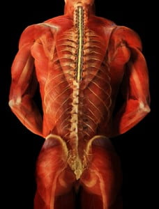
degenerative disc disease Archives

Sitting with your legs crossed may be contributing to your back pain

Think of some of the ways you sit. Whether taking a ride on the subway, waiting patiently at the doctor’s office, or sitting as a passenger in a car, the way we sit, especially for prolonged periods can have an effect on the pain we may experience in our low back.


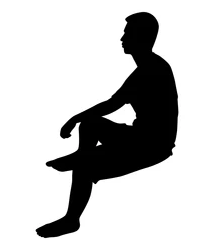
For many of us, sitting with our legs crossed feels comfortable in the moment but have you ever considered how this can affect your back and cause pain? What happens to your spine and muscles when sitting with your legs crossed for an extended period of time? When considering good posture, a large component is keeping your spine neutral and avoiding positions that twist the spine or cause misalignment. When sitting with your legs crossed, your pelvis becomes tilted and unconsciously, we tilt our entire torso towards one side. There is a curvature towards one side in the low back which then creates problems in other areas of the spine. Naturally, with a tilt of the pelvis, the upper body will compensate by leaning the opposite direction to maintain balance. Working from the bottom up, pressure on the lumbar spine will cause curvature and misalignment in the thoracic spine. This places strain on the cervical spine to keep the head in the upright position.


Sitting with our legs crossed can also affect our muscles. There are two common sitting positions that we should be mindful of, knee over knee or foot over knee for a prolonged period can cause a tightness in the hamstrings, hip flexors and glute muscles. The iliopsoas, one of the major hip flexor muscles, is responsible for external rotation of the femur and an integral part in maintaining posture. This muscle may become strained due to contraction while the pelvis is tilted. You may notice soreness and tenderness in the low back and hip area. Similarly, sitting foot over knee can over time damage the muscles in the inner thigh. Most commonly, the sartorius muscle can become tight and cause discomfort when sitting with the calf over the knee. At the Norman Marcus Pain Institute, Iliacus and Psoas muscles on the hip can be treated for pain. As one of the most common muscles causing pain for patients, when treated, the Iliacus and other hip flexors can provide great relief for pain in the low back, buttock and groin.

Instead of sitting with crossed legs, sit in a chair with height necessary to place both feet flat on the floor. While sitting upright with your back against the chair, you may want to consider placing a cushion under your bottom that can add support to the low back.

References

Lee BJ, Cha HG, Lee WH. The effects of sitting with the right leg crossed on the trunk length and pelvic torsion of healthy individuals. J Phys Ther Sci. 2016 Nov;28(11):3162-3164. doi: 10.1589/jpts.28.3162. Epub 2016 Nov 29. PMID: 27942141; PMCID: PMC5140821.

Share

In today’s world we are bombarded by information and ways to obtain it. Do you want to know how many steps you take a day? Just buy a device and it will calculate it for you. Care to know what your cholesterol levels are? Simply go get a kit at your nearest drug store. Is it possible we are gathering too much information for our own good?

The same question can be asked about medical testing. Is it useful or even helpful to know certain things about our bodies? Many of us have undoubtedly had the experience where we have gone to the doctor because we had pain and an imaging study was ordered. MRI, CT, and ultrasound can give us information about our bodies that would have been unimaginable in the past.

While technology in general is of course a huge benefit, at the Norman Marcus Pain Institute, we believe it is necessary to proceed with caution as we navigate through it all. At NMPI, one common problem we find in back pain, neck pain, and shoulder pain is the overuse and overreliance on imaging studies. “Abnormalities” are often found on imaging studies without any relation to one’s pain. In fact, seeing abnormalities is more common than seeing a “normal” spine!   For example, up to 40% of people without back pain can haveBack Pain herniated discs and as many as 70% may have degenerated discs. That is why the US Institute of Medicine suggests not getting an MRI too quickly since you are likely to see one of these problems and assume it is the cause of your pain. This can lead to unnecessary nerve blocks and surgery.

At NMPI, we often see patients who have a herniated disc, yet they only have pain in their back and buttock. They have not experienced any radiation into the leg(s) at all. In these patients, the pain generally has nothing to do with the disc herniation since disc herniation pain generally radiates down the leg and into the foot. Even when the patient is experiencing pain down the leg with a herniated disc found on MRI, muscles in the low back and buttocks may be the cause of the pain. If muscles are not examined as a potential cause, you may undergo an apparently reasonable surgery, without achieving relief of pain. Some studies show that up to 50% of spine surgeries fail (resulting in failed back surgery syndrome) and one of the reasons is the failure to identify muscles that were the true source of the pain.

Most back pain and neck pain is caused by soft tissue such as muscles and tendons. This is confusing because you may have been told your pain is from your spine, discs, or nerves. The problem is most people as they get older have signs of wear and tear on their x-rays and MRIs, but these common signs of aging may not explain your pain if the pain actually originates in your muscles. That’s why we say, when diagnosing persistent pain it’s not having “more” information at hand, it’s having the “right” information at hand.

At NMPI, we often see patients who experience persistent pain even after multiple spine surgeries. Our non-surgical, non-invasive treatment program has most of our patients leaving our office free of long standing back, neck, shoulder, and headache pain.

Share

Earlier in November, Rafael Nadal, the 14-time Grand Slam winner, announced he would receive stem cell treatment to help heal his ailing back, the same type of treatment he received for his knee. His doctor in Barcelona, Dr. Angel Ruiz-Cotorro, who has been treating Nadal for 14 years, said, “Nadal’s back pain is ‘typical of tennis’ players in that the treatment is meant to help repair his cartilage.” Stem cells were recently extracted from Nadal for a cultivation process to “produce the necessary quantities,” said Ruiz-Cotorro. Once cultivated, the stem cells will be placed into the joints of his spine with the goal of regeneration of cartilage as well as for an anti-inflammatory effect. Dr. Ruiz-Cotorro predicts that Nadal can return to training in early December.

Will stem cell treatment work for Nadal’s Back Pain?

Stem cell treatment may seem logical in certain situations – for example, if you have a mechanical problem where a knee has worn out cartilage, causing bone to rub against bone, it makes sense to use stem cells to grow new cartilage to have a cushion to protect the bone and cause the knee to be less painful. As much as we may want to see him back on the courts tennisgrabbing more grand slam titles, if Nadal’s stem cell treatment is being used to eliminate his pain by repairing his joints or discs, the actual cause of his back pain may not be addressed.
Where does back pain originate?

The number one reason for back pain is muscular and other soft tissue, yet muscles are rarely evaluated as the cause of back pain. The only way to determine if Nadal’s back pain is from soft tissue and similar to most people with back pain would be a physical examination of Nadal’s back that included identifying possible muscles as the cause of his pain.

Some doctors believe that the disc, the cushion between the bones of the spine (the vertebra), is a major cause of back pain. They believe that surgeries to correct the flattening or herniation of the disc will decrease or eliminate back pain. Sometimes they are right, but they are just as likely to be wrong. The truth is that there is as high as a 50% failure rate for spine surgeries that were done to eliminate back pain thought to be related to disc problems. There are other joints in the spine that are thought to cause pain; one of them is the facet joint, which could also be a target for stem cell treatments.

When doctors rely on an MRI or CT scan to determine the source of the pain, the information obtained is often confusing. If a surgeon sees an abnormality on an MRI, he will often point to that abnormality as the cause of the pain; in my experience the abnormality found on an MRI or CT scan frequently is not the cause. In fact, if you randomly selected 100 people off the street, and perform an imaging scan, 40 may present with a herniated disc and have no pain and absolutely no awareness of their herniated disc; 70 may have degenerated (worn) discs with no pain, and a large number will have facet joint abnormalities. Therefore, finding an “abnormality” is more common than not. One, then, can deduce that the abnormality is more likely NOT the source of the pain. So treating the abnormality (with steroid injections, surgery, or stem cells) may therefore not relieve the pain.

Stem Cell Treatment and Sports Stars

Nadal, currently ranked as the number 3 professional tennis player in the world, is not the first sports star to chase after a “miracle cure.” The Denver Bronco quarterback Payton Manning and Yankees pitcher Bartolo Colon both went abroad to seek out stem cell treatment as a quick fix to get back in the game. (They both seem to be doing better overall, but it is inconclusive if the stem cell treatment was the cause of their recovery).

Will it work?

In the laboratory, it has been possible to demonstrate the ability of stem cells (most commonly found in the developing embryo and newborn) to grow new tissue. These cells are like silly putty; they can turn into, or adapt, to become any type of tissue. For example, a stem cell in the right environment in the body could become bone, cartilage or some organ (for example, liver or pancreas). But, it hasn’t been as easy to grow tissue in an actual person. There are some early studies that show that stem cells “may” relieve back pain, but both the doctors who are testing the technique and outside experts say much more research is needed before they can say whether the treatment offers real relief.

The use of stem cell therapies continues to be a hot topic for debate in the sports medicine and orthopedic surgery worlds. There is no current evidence-based research to prove that it works.

Sir William Osler, a famous physician, once said: “Use every new treatment as quickly as possible before it stops working.” Stem cell treatment needs to be further investigated to determine if stem cell treatments indeed work, and if so, for what conditions?


Share

Muscles as the source of pain

Muscles are the largest organ system in the body, accounting for approximately 50% of our body weight.  We have different ways of categorizing muscles: how they look, how they move, and where they’re located. We are going to focus on muscles that move voluntarily, which means we tell the muscles to move, as opposed to involuntary muscles, which automatically move on their own (like our heart and blood vessels). There are 641 muscles in the body – 340 pairs (meaning we have one on the right, and one on the left), and one unpaired (which is the transverse arytenoid for those who must know).

Back Pain

70% of lower back pain is diagnosed as idiopathic or non-specific[1], which means we are not sure what caused the pain. However, most investigators believe that sprains and strains of the soft tissue are the source of pain. Soft tissue refers to muscles, tendons, and ligaments. So it may be surprising that the emphasis in evaluating and treating lower back pain, neck pain, and shoulder pain, is on the spine and the nerves coming out of the spine. In fact, from 1997 to 2005, the prevalence of the diagnosis of spine-related issues has increased 100% while the diagnosis of strains and sprains of soft tissue has gone down by 40%[2].

This is generally attributed to the increase in the use of high-tech imaging studies, such as MRI and CT scans.  However, just because we have a clearer image of what’s going on inside of your body doesn’t mean that we have a clearer understanding of what’s causing your pain. More than 90% of lower spine MRIs exams in adults are abnormal[3]. Studies have found that up to 40% of people have herniated discs and as many as 70% have degenerated discs with no pain. If people can walk around with abnormal spines without pain, then this means that abnormalities in the spine aren’t always the cause of pain. Your diagnosis of a herniated disc, spinal stenosis, or spondylosisthesis may actually be unrelated to your source of pain.

Muscles are often ignored when it comes to diagnosing pain. I believe the reason is that we rarely evaluate muscles as a source of pain. We generally don’t learn about or understand how they work, what chemical changes take place inside, and how they produce pain (the pathophysiology). I would like to take the next few blogs to discuss how muscles contribute to your chronic pain.


[1] Deyo, RA., et al. Low Back Pain. NEJM. 2001; 344(5):363-370

[2] Martin, B., et al. Expenditures and health status among adults with back and neck problems. JAMA. 299(6):656-64, Feb 2008.

[3] Zimmerman, Robert D. “A Review of Utilization of Diagnostic Imaging in the Evaluation of Patients with Back Pain: The When and What of Back Pain Imaging.” Journal of Back and Musculoskeletal Rehabilitation 8 (1997): 125-33.

Share

Apparent diagnoses sometimes lead us astray …

When treating chronic back pain, over-reliance on imaging studies frequently suggest arthritis, disc herniations, spinal stenosis, degenerative disc disease, and spondylolisthesis as the underlying diagnoses. Unsuccessful treatment often results if muscles, tendons, and fascia are not considered in the differential diagnosis.

Widespread pain is often diagnosed as Fibromyalgia Syndrome (FMS). In 1990 the American College of Rheumatology (ACR) presented classification criteria for the diagnosis of FMS [http://bit.ly/1bCzlC4.] 11 of 18 specified tender points as well as widespread pain defined as pain experienced on the left and right side, in the upper and lower body, and axially. Most patients are diagnosed with FMS by their primary care physician. However, most primary care physicians do not perform a physical examination of tender points. Because of this the ACR in 2010 proposed an additional set of diagnostic criteria for FMS that did not rely on a physical examination (http://bit.ly/1bCzlC4).

Three conditions must be met to satisfy the criteria:

  1. Threshold scores on two new indices created by the ACR- the widespread pain index and the symptom severity scale score based on the presence of fatigue, waking un-refreshed and cognitive symptoms.
  2. Patients must have similar symptoms at approximately the same intensity for at least three months.
  3. No other disorder would reasonably explain the pain.

Although there is reasonable correlation between patients diagnosed with the 1990 criteria and the 2010 criteria, the absence of the physical examination may lead to overlooking patients whose diffuse pain can be successfully treated by addressing peripheral pain generators. Painful tissue peripherally (for example muscle or joint) can sensitize muscles diffusely [Woolf CJ, Central Sensitization: Implications for the diagnosis and treatment of pain. Pain 152 (2011) S2-S 15].

I welcome your comments on the complexities of diagnosing and treating FMS; more on central sensitization in my next post.


Share
NORMAN MARCUS PAIN INSTITUTE
30 East 40th Street - New York, NY 10016
Tel 212-532-7999 Fax 212-532-5957
Share
Help Desk Software