Spinal stenosis Archives

Sitting with your legs crossed may be contributing to your back pain

Think of some of the ways you sit. Whether taking a ride on the subway, waiting patiently at the doctor’s office, or sitting as a passenger in a car, the way we sit, especially for prolonged periods can have an effect on the pain we may experience in our low back.


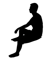
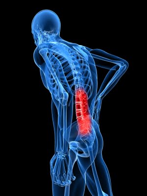
For many of us, sitting with our legs crossed feels comfortable in the moment but have you ever considered how this can affect your back and cause pain? What happens to your spine and muscles when sitting with your legs crossed for an extended period of time? When considering good posture, a large component is keeping your spine neutral and avoiding positions that twist the spine or cause misalignment. When sitting with your legs crossed, your pelvis becomes tilted and unconsciously, we tilt our entire torso towards one side. There is a curvature towards one side in the low back which then creates problems in other areas of the spine. Naturally, with a tilt of the pelvis, the upper body will compensate by leaning the opposite direction to maintain balance. Working from the bottom up, pressure on the lumbar spine will cause curvature and misalignment in the thoracic spine. This places strain on the cervical spine to keep the head in the upright position.


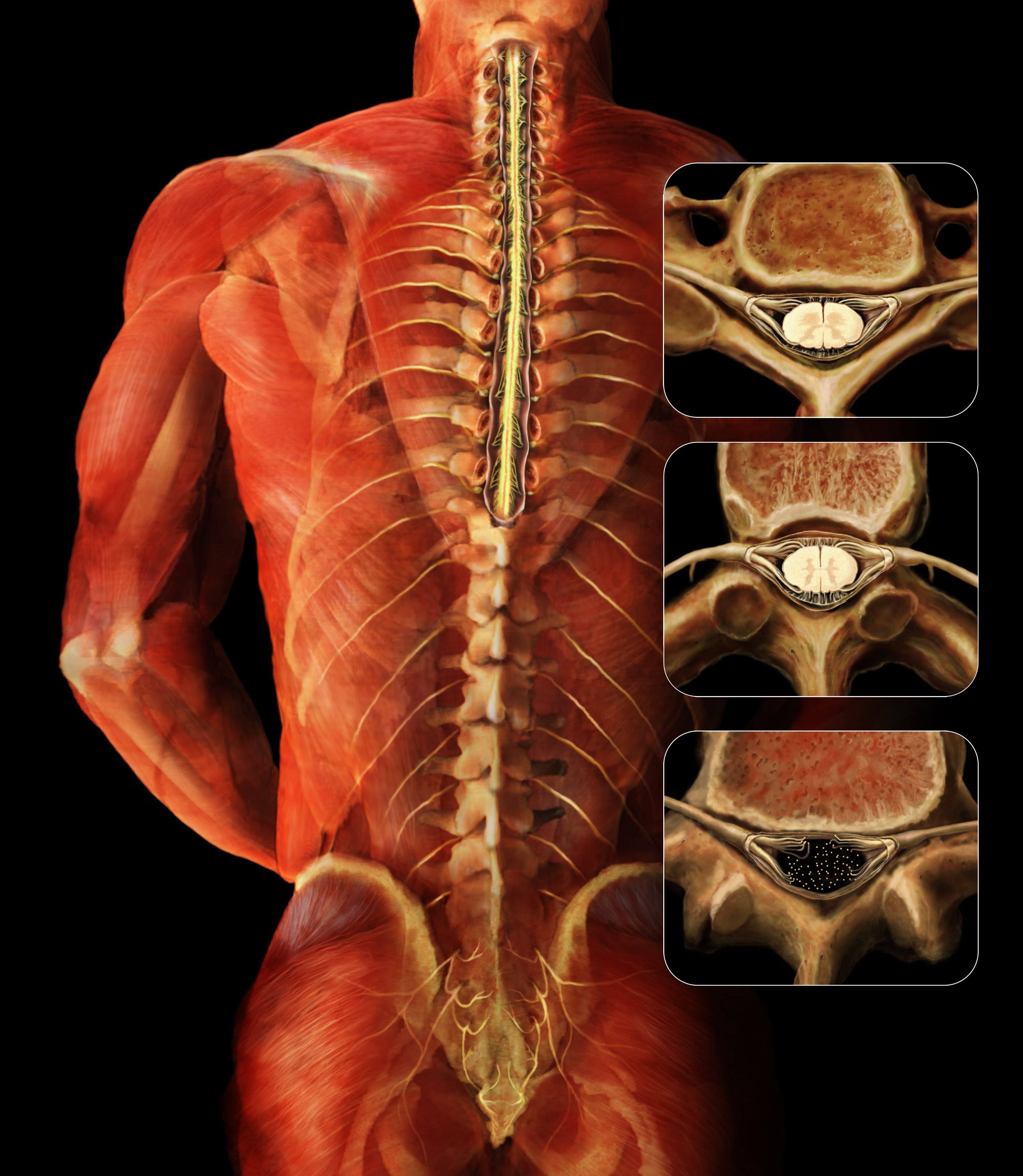
Sitting with our legs crossed can also affect our muscles. There are two common sitting positions that we should be mindful of, knee over knee or foot over knee for a prolonged period can cause a tightness in the hamstrings, hip flexors and glute muscles. The iliopsoas, one of the major hip flexor muscles, is responsible for external rotation of the femur and an integral part in maintaining posture. This muscle may become strained due to contraction while the pelvis is tilted. You may notice soreness and tenderness in the low back and hip area. Similarly, sitting foot over knee can over time damage the muscles in the inner thigh. Most commonly, the sartorius muscle can become tight and cause discomfort when sitting with the calf over the knee. At the Norman Marcus Pain Institute, Iliacus and Psoas muscles on the hip can be treated for pain. As one of the most common muscles causing pain for patients, when treated, the Iliacus and other hip flexors can provide great relief for pain in the low back, buttock and groin.

Instead of sitting with crossed legs, sit in a chair with height necessary to place both feet flat on the floor. While sitting upright with your back against the chair, you may want to consider placing a cushion under your bottom that can add support to the low back.

References

Lee BJ, Cha HG, Lee WH. The effects of sitting with the right leg crossed on the trunk length and pelvic torsion of healthy individuals. J Phys Ther Sci. 2016 Nov;28(11):3162-3164. doi: 10.1589/jpts.28.3162. Epub 2016 Nov 29. PMID: 27942141; PMCID: PMC5140821.

Share

Spinal Surgery for Lumbar Stenosis

A recent study was published in the Journal of the American Medical Association, analyzing spinal surgeries done on Medicare patients who were diagnosed with lumbar stenosis.

Let’s rewind – what is lumbar stenosis? Lumbar refers to the lower part of the spine, where we would normally identify as low back. Stenosis refers to the narrowing of the bones in the spine, often pushing or compressing the nerves that being in the spinal cord and extend into your legs. Spinal stenosis is often associated with pain and numbness in the leg.

When a patient is diagnosed with lumbar spinal stenosis, a wide variety of treatments are offered. If the spinal stenosis is actually causing pain and difficulties functioning, such as walking, you would imagine that exercise and physical therapy would have little or no effect. However, pBack Painhysical therapy and exercise are frequently prescribed and may reduce symptoms. This means that at least some of the pain is from muscles and that the picture seen with the CT or MRI showing the nerves being compressed wasn’t accurately showing the reason for all the pain. Without a thorough examination of the muscles in the lower body, we may overlook an easily treated source of pain.

Overreliance on imaging studies can lead to unnecessary, costly, and sometimes damaging treatments, which may include various pain medications, injections (such as epidural injections), and surgery. When surgery is recommended, there are different complexities of surgeries that can be performed.

In this recent study, the surgeries were divided into three categories: 1. Decompression by itself (removing a small piece of bone from the spine to relieve pressure on the spinal cord or nerves), 2. Simple (only 1-2 levels) fusion (fusing the bones in the spine using metal and or bone that may be harvested from the hip or provided by a bone bank), or 3. Complex Fusion (more than 2 levels and/or anterior [front] and posterior [back] approach). From the years 2002 to 2007, the number of decompression and simple fusion surgeries decreased while the number of complex fusion surgeries increased 15-fold. With increasing complexity of the surgery, the odds for complications and rehospitalization significantly increase without a significant demonstrated improvement in outcomes.

We should all be concerned that the number of complex surgeries is increasing, causing more severe side effects and death. Hospital charges alone for a less complex surgery can cost around $23,000 while the complex surgeries may cost over $80,000.


Share

In today’s world we are bombarded by information and ways to obtain it. Do you want to know how many steps you take a day? Just buy a device and it will calculate it for you. Care to know what your cholesterol levels are? Simply go get a kit at your nearest drug store. Is it possible we are gathering too much information for our own good?

The same question can be asked about medical testing. Is it useful or even helpful to know certain things about our bodies? Many of us have undoubtedly had the experience where we have gone to the doctor because we had pain and an imaging study was ordered. MRI, CT, and ultrasound can give us information about our bodies that would have been unimaginable in the past.

While technology in general is of course a huge benefit, at the Norman Marcus Pain Institute, we believe it is necessary to proceed with caution as we navigate through it all. At NMPI, one common problem we find in back pain, neck pain, and shoulder pain is the overuse and overreliance on imaging studies. “Abnormalities” are often found on imaging studies without any relation to one’s pain. In fact, seeing abnormalities is more common than seeing a “normal” spine!   For example, up to 40% of people without back pain can haveBack Pain herniated discs and as many as 70% may have degenerated discs. That is why the US Institute of Medicine suggests not getting an MRI too quickly since you are likely to see one of these problems and assume it is the cause of your pain. This can lead to unnecessary nerve blocks and surgery.

At NMPI, we often see patients who have a herniated disc, yet they only have pain in their back and buttock. They have not experienced any radiation into the leg(s) at all. In these patients, the pain generally has nothing to do with the disc herniation since disc herniation pain generally radiates down the leg and into the foot. Even when the patient is experiencing pain down the leg with a herniated disc found on MRI, muscles in the low back and buttocks may be the cause of the pain. If muscles are not examined as a potential cause, you may undergo an apparently reasonable surgery, without achieving relief of pain. Some studies show that up to 50% of spine surgeries fail (resulting in failed back surgery syndrome) and one of the reasons is the failure to identify muscles that were the true source of the pain.

Most back pain and neck pain is caused by soft tissue such as muscles and tendons. This is confusing because you may have been told your pain is from your spine, discs, or nerves. The problem is most people as they get older have signs of wear and tear on their x-rays and MRIs, but these common signs of aging may not explain your pain if the pain actually originates in your muscles. That’s why we say, when diagnosing persistent pain it’s not having “more” information at hand, it’s having the “right” information at hand.

At NMPI, we often see patients who experience persistent pain even after multiple spine surgeries. Our non-surgical, non-invasive treatment program has most of our patients leaving our office free of long standing back, neck, shoulder, and headache pain.

Share

Epidural steroids are not effective for spinal stenosis

I have been suggesting for many years that there is an overuse of spinal injections and surgeries for low back and leg pain, so it was no surprise when I read an article in The New York Times (NYT) that reported on a study recently published in the New England Journal of Medicine[1] about patients with spinal stenosis who are frequently treated with a procedure that has been shown to be ineffective. Epidural steroid and lidocaine injections for patients found to have spinal stenosis were no better than epidural injections of lidocaine alone. In chapter 4 of my book End Back Pain Forever I discuss the many reasons for back pain. MRIs and CT scans of the low back will almost always find something such as spinal stenosis or a degenerated or herniated disc even in patients without pain. Therefore the US Institute of Medicine suggests that these imaging studies should not be done routinely since what you find in the study is frequently not the cause of your pain. The most common cause of back pain is muscles and other soft tissue. If you treat the diagnosis you got from the MRI or CT and the actual reason for the pain is muscle, it makes sense that the treatment will frequently fail, which it does!

The NYT report of this relatively large, randomized, double blind, controlled study clearly demonstrates the ineffectiveness of the use of steroids for symptoms attributable to spinal stenosis (narrowing of the canal in the spine which contains the spinal cord) which occurs frequently as we age. The treatments most often provided are epidural steroids to theoretically reduce the inflammation of the nerves being squeezed by the narrow canal, and surgery to widen the canal. Both approaches have a significant failure rate.

At the Norman Marcus Pain Institute we have shown in multiple published articles that one reason why these approaches are ineffective is that the pain in the back and leg may not be the result of the narrowing or other supposed abnormalities seen on the MRI or CT scan. It is well known that very few (in one published article- less than 10%) scans of the low back in adults are read as normal; as many as 40% of adults with no back pain have herniated discs, and 70% have degenerated discs. So it is “normal” to find an abnormality.

B_vertebrae_function_01

Our unique physical examination, utilizes an instrument I developed, reveals that many patients with back and leg pains have areas of muscle tenderness that are the source of their pain yet are overlooked. One study of more than 23,000 patients[2] found that 70-80% of patients with back pain were diagnosed with sprains and strains of muscle and other soft tissue. It’s hard to believe then, armed with this knowledge, that muscle examination and treatment is not part of the typical standard of care for back pain in the USA[3]. If we are to properly address the cause of most back pain, the evaluation and treatment of its muscular causes must be addressed.


[1] Friedly JL, Comstock BA, Turner JA, et al. A Randomized Trial of Epidural Glucocorticoid Injections for Spinal Stenosis. New England Journal of Medicine. 2014:374:11-21.

[2] Deyo RA, Weinstein JN. Primary care – low back pain. New England Journal of Medicine. 2001:5:363-70.

[3] Chou R, Qaseem A, Snow V, Casey D, Cross JT, Shekelle P, et al. Diagnosis and Treatment of Low Back Pain: A Joint Clinical Practice Guideline from the American College of Physicians and the American Pain Society. Ann Intern Med. 2007;147:478-491.

Share

Muscles as the source of pain

Muscles are the largest organ system in the body, accounting for approximately 50% of our body weight.  We have different ways of categorizing muscles: how they look, how they move, and where they’re located. We are going to focus on muscles that move voluntarily, which means we tell the muscles to move, as opposed to involuntary muscles, which automatically move on their own (like our heart and blood vessels). There are 641 muscles in the body – 340 pairs (meaning we have one on the right, and one on the left), and one unpaired (which is the transverse arytenoid for those who must know).

Back Pain

70% of lower back pain is diagnosed as idiopathic or non-specific[1], which means we are not sure what caused the pain. However, most investigators believe that sprains and strains of the soft tissue are the source of pain. Soft tissue refers to muscles, tendons, and ligaments. So it may be surprising that the emphasis in evaluating and treating lower back pain, neck pain, and shoulder pain, is on the spine and the nerves coming out of the spine. In fact, from 1997 to 2005, the prevalence of the diagnosis of spine-related issues has increased 100% while the diagnosis of strains and sprains of soft tissue has gone down by 40%[2].

This is generally attributed to the increase in the use of high-tech imaging studies, such as MRI and CT scans.  However, just because we have a clearer image of what’s going on inside of your body doesn’t mean that we have a clearer understanding of what’s causing your pain. More than 90% of lower spine MRIs exams in adults are abnormal[3]. Studies have found that up to 40% of people have herniated discs and as many as 70% have degenerated discs with no pain. If people can walk around with abnormal spines without pain, then this means that abnormalities in the spine aren’t always the cause of pain. Your diagnosis of a herniated disc, spinal stenosis, or spondylosisthesis may actually be unrelated to your source of pain.

Muscles are often ignored when it comes to diagnosing pain. I believe the reason is that we rarely evaluate muscles as a source of pain. We generally don’t learn about or understand how they work, what chemical changes take place inside, and how they produce pain (the pathophysiology). I would like to take the next few blogs to discuss how muscles contribute to your chronic pain.


[1] Deyo, RA., et al. Low Back Pain. NEJM. 2001; 344(5):363-370

[2] Martin, B., et al. Expenditures and health status among adults with back and neck problems. JAMA. 299(6):656-64, Feb 2008.

[3] Zimmerman, Robert D. “A Review of Utilization of Diagnostic Imaging in the Evaluation of Patients with Back Pain: The When and What of Back Pain Imaging.” Journal of Back and Musculoskeletal Rehabilitation 8 (1997): 125-33.

Share

Physical Examinations Result in Better Outcomes for Back Pain

Donna, a 43 year old married mother with a one year old son saw me for evaluation of severe low back and buttock pain. Her MRIs showed a disc herniation at L4-5 and moderate-to-severe spinal stenosis at the same level. She received physical therapy, 3 epidural steroids and chiropractic treatments with transient or no pain relief. Her orthopedic surgeon told her that since all conservative measures were already done the only option was spine surgery. He felt a fusion should be done.

My soft tissue examination identified 3 areas in her low back (the Quadratus Lumborum on both sides, and the right Gluteus Maximus) as possible sources of her pain. Each was treated with an injection technique that emphasizes placement of the needle into the muscle’s tendinous and bony attachments and the tissue along the course of the muscle from the origin to the insertion. A 3 day post-injection physical therapy protocol followed each injection session. Donna was taught all 21 exercises in my low back exercise program and experienced complete relief in less than a month after starting treatment. She returned to the gym, ran a half marathon and on follow up 5 years after treatment, was still pain free, hiking, biking, and running.blog post from NMPI

I believe that patients like Donna who receive spine surgery will frequently be found as Failed Back Surgery Syndrome cases.

Imaging findings often do not provide an accurate explanation for your pain. Addressing the imaging diagnosis without an examination to identify possible specific sources of muscle pain may lead to treatments that are at best inadequate and at worst damaging.

Share

A step care model for pain management is often the best choice

James was a 67-year-old, married entrepreneur with bilateral thigh and calf pain that prevented him from walking for more than one block on a flat surface or sitting for more than 30 minutes. He couldn’t leave his house for a walk and was unable to sit in a restaurant. His pain was 7/10.  I evaluated him after he had undergone 2 lumbar spine fusions, epidural steroid and trigger point injections, all without pain relief.  His history revealed that after a fall he developed lower back and leg pain. An MRI found spinal stenosis and degenerative spondylolisthesis.  He had  a spinal fusion, which provided 2 months of relief before all of his symptoms returned.  He tried trigger point injections and epidural steroids, and then a second spinal fusion – all of which provided no lasting relief.Norman Marcus Pain Institute-back-pain

When James came to see me, he was offered an indwelling morphine pump or a spinal cord stimulator.

His primary complaint was a constant burning and pulling sensation in his thighs, and a pain that shot down the back of his leg to his heels. On examination, I discovered James was deconditioned: he had decreased range of motion in his back and hips due to stiffness, and showed weakness in his abdominals and back extensor muscles.  James’ physical examination also revealed 4 muscles that were likely contributing to his pain:  the right and left gluteus maximus, the tensor fasciae latae, and the vastus lateralis.  He received muscle-tendon injections to each muscle, and following each procedure,  3 days of a structured physical therapy protocol.

Soon after receiving all injections and learning all 21 exercises, he was walking easily on the street, eating in restaurants, and was able to travel to Vietnam and China with his wife.

His imaging findings of stenosis and spondylolisthesis existed before his fall and did not produce symptoms. He only had back and leg pain after his fall. It would have made sense to consider that soft tissue injury was a reasonable possible source of his pain prior to embarking on costly, interventions with considerable downside risks. I am suggesting that a step care model would have been a better option for James and for all of our patients with back pain (simple and cheap before complicated and expensive).

Share

Use of statins may be cause of weakness in legs

Norman Marcus Pain Institute-blog-Nov12I have a friend who has been complaining of weakness in his legs. He has a history of high cholesterol and has been on statins.

He was seen by a spine surgeon and after an MRI showed spinal stenosis, decompressive surgery was suggested. His internist recommended stopping the statin since it can cause leg pain and weakness. Two weeks after the statin was stopped his weakness went away.

Overreliance on imaging studies could result in unnecessary surgeries as in this case. As many as 90% of adult patients who have had MRIs of the lumbosacral spine have spinal “abnormalities”. Just because we find something on imaging doesn’t mean it is the cause of the pain.

Share

Apparent diagnoses sometimes lead us astray …

When treating chronic back pain, over-reliance on imaging studies frequently suggest arthritis, disc herniations, spinal stenosis, degenerative disc disease, and spondylolisthesis as the underlying diagnoses. Unsuccessful treatment often results if muscles, tendons, and fascia are not considered in the differential diagnosis.

Widespread pain is often diagnosed as Fibromyalgia Syndrome (FMS). In 1990 the American College of Rheumatology (ACR) presented classification criteria for the diagnosis of FMS [http://bit.ly/1bCzlC4.] 11 of 18 specified tender points as well as widespread pain defined as pain experienced on the left and right side, in the upper and lower body, and axially. Most patients are diagnosed with FMS by their primary care physician. However, most primary care physicians do not perform a physical examination of tender points. Because of this the ACR in 2010 proposed an additional set of diagnostic criteria for FMS that did not rely on a physical examination (http://bit.ly/1bCzlC4).

Three conditions must be met to satisfy the criteria:

  1. Threshold scores on two new indices created by the ACR- the widespread pain index and the symptom severity scale score based on the presence of fatigue, waking un-refreshed and cognitive symptoms.
  2. Patients must have similar symptoms at approximately the same intensity for at least three months.
  3. No other disorder would reasonably explain the pain.

Although there is reasonable correlation between patients diagnosed with the 1990 criteria and the 2010 criteria, the absence of the physical examination may lead to overlooking patients whose diffuse pain can be successfully treated by addressing peripheral pain generators. Painful tissue peripherally (for example muscle or joint) can sensitize muscles diffusely [Woolf CJ, Central Sensitization: Implications for the diagnosis and treatment of pain. Pain 152 (2011) S2-S 15].

I welcome your comments on the complexities of diagnosing and treating FMS; more on central sensitization in my next post.


Share

I have heard from a number of group members who have been diagnosed with spinal stenosis. The diagnosis is often made based on what is found on a CT scan or MRI without the expected corresponding signs and symptoms. It is important to understand what any diagnosis means in relationship to your back or leg pain.

Spinal stenosis is a narrowing of the space formed by the bony segments that together make up the vertebra (the bones of the spine). The space in each vertebra is connected to the vertebra above and below to form the spinal canal, through which the spinal cord passes. Narrowing of the canal in the lumbar region, called lumbar spinal stenosis, could squeeze the spinal cord. When you are standing up, the curve in the spine makes the narrowing worse and may cause pain radiating to the leg. Many patients found to have narrowing don’t have the signs and symptoms that would indicate that their back and leg pain was caused by the narrowing. Bending over when you walk, having more pain if you straighten up, and having to wait a few minutes when you sit down for the pain to go away, are all symptoms that suggest the spinal stenosis was truly the cause of the pain; just finding narrowing with imaging isn’t enough.

Other imaging diagnoses such as degenerative disc disease, degenerative osteoarthritis, bulging or herniated disc, and facet arthropathy, may also be misleading. Just because there is an anatomic finding on an image doesn’t mean it is the cause of the pain. If some form of exercise relieved the pain, the most reasonable explanation would be that much of the pain was related to soft tissue, such as muscle and tendon and not to the imaging diagnoses.


Share
NORMAN MARCUS PAIN INSTITUTE
30 East 40th Street - New York, NY 10016
Tel 212-532-7999 Fax 212-532-5957
Share
Help Desk Software